Schedule Your Free Consultation

The tools a dentist uses directly affect the accuracy of the diagnosis, theprecision of the treatment, and the quality of the result. At Michael Kriston DDS in Danville, we’ve invested in the latest imaging, scanning, diagnostic,and treatment technology — not because it’s impressive to have, but because it makes your care measurably better.

Every piece of technology in our office was selected for a specific clinical purpose.Our 3D imaging reveals anatomy that traditional X-rays can’t show. Our digital scanner eliminates the discomfort of impression trays. Our AI-assisted diagnostic tools provide a second set of “eyes” that enhance Dr. Kriston’s 38 years of clinical experience. And our dental laser makes soft tissue procedures more precise, more comfortable, and faster to heal.

Our imaging and scanning equipment — the Care stream 3D CBCT, the Medit intraoral scanner, and the Care stream intraoral cameras — were all purchased brand new in 2025, ensuring you benefit from the most current technology available.

Contact

Carestream CS 8200 3D — Cone Beam CT Imaging

Traditional dental X-rays give a two-dimensional view of a three-dimensional structure.They’re valuable for many purposes, but for implant planning they have seriouslimitations — overlapping anatomy, no depth information, no ability to visualize bone density or nerve pathways, and inherent distortion that makes measurements unreliable. A dentist attempting to plan implant placement from a two-dimensional film is not seeing the full picture of the area, and the measurements they’re working from may not be accurate.

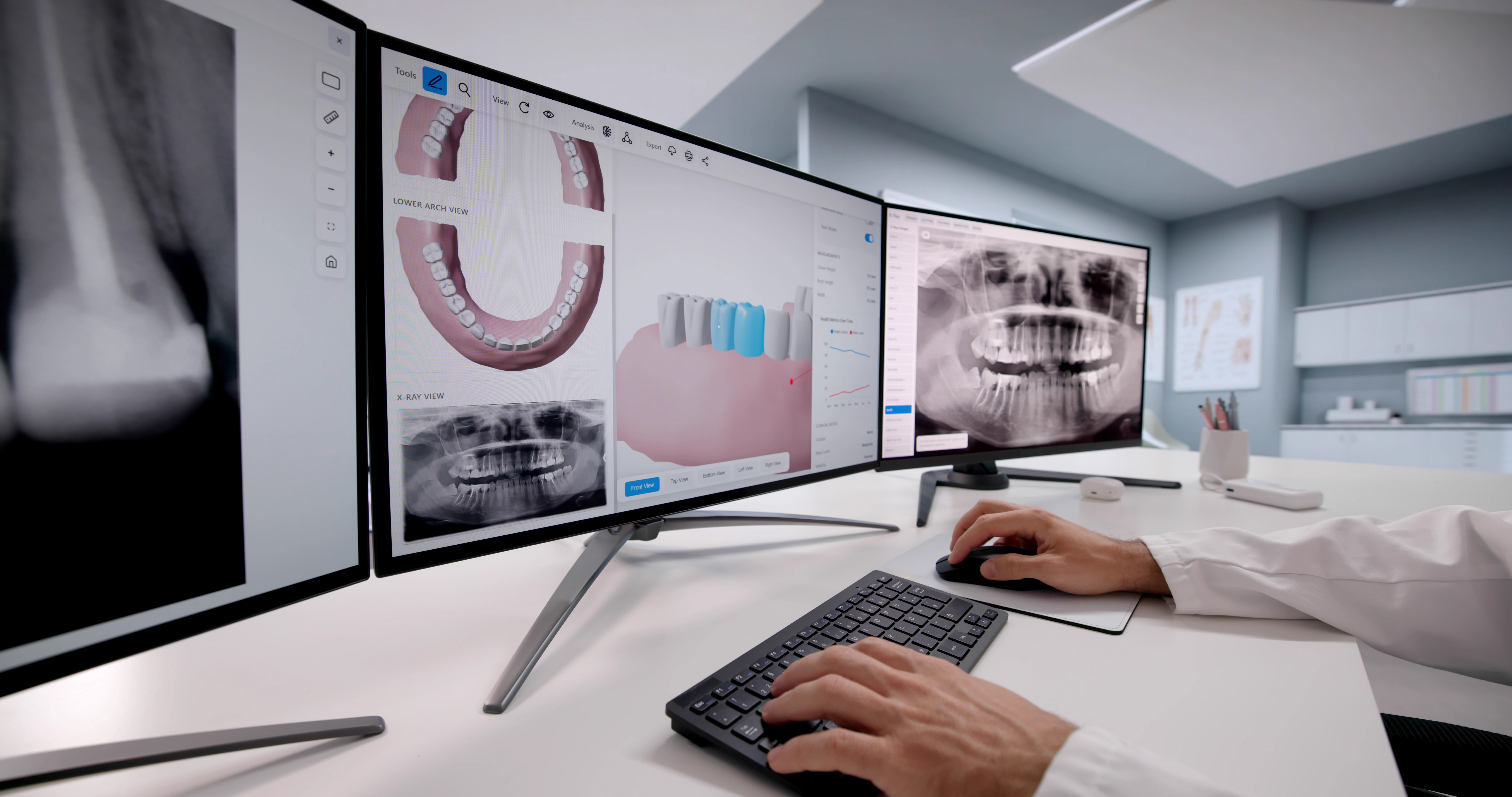
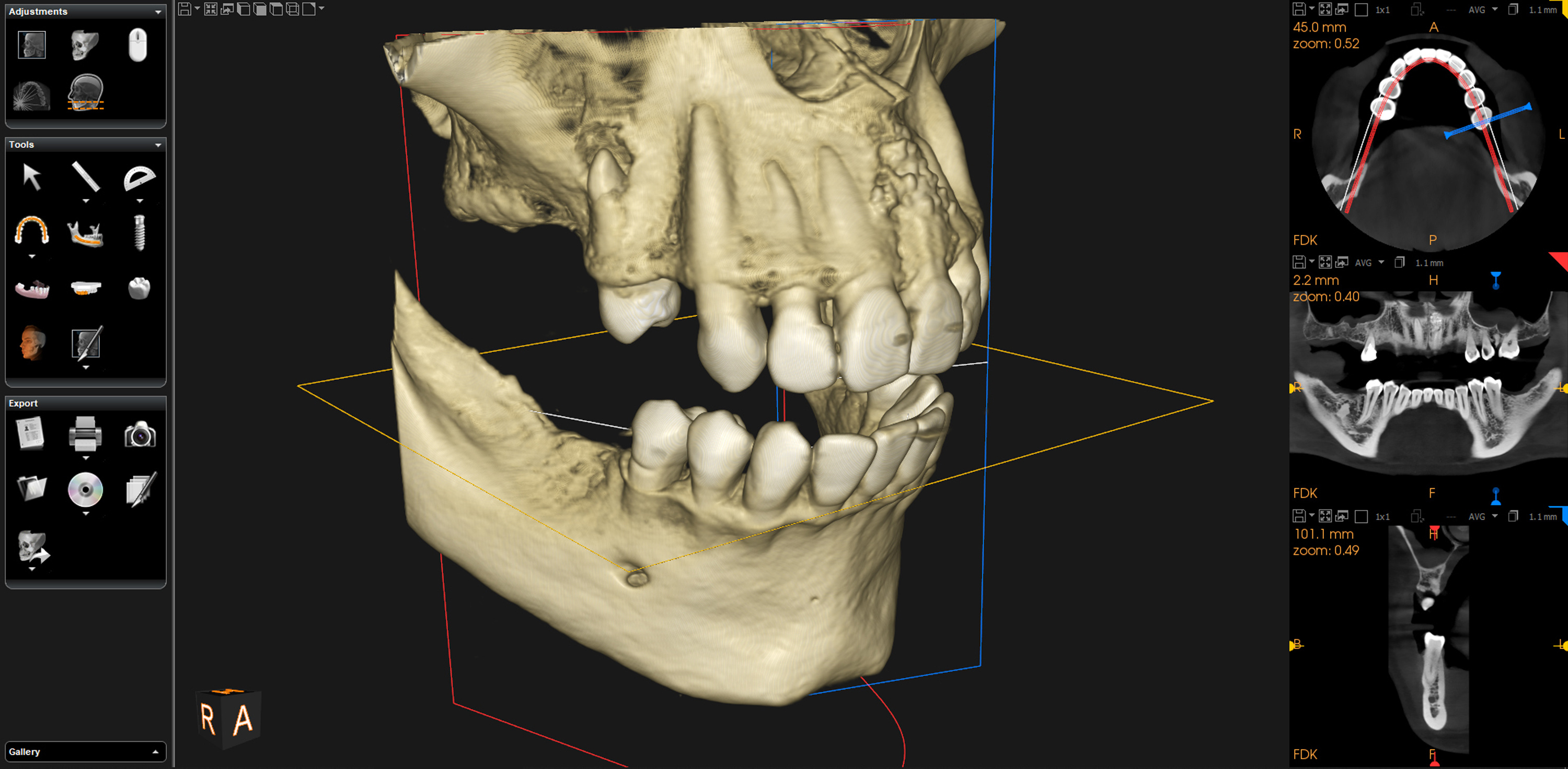
This is why 3D CBCT imaging is now the gold standard for dental implant placement. Our Care stream CS 8200 3D cone beam CT scanner produces a detailed, undistorted 3D volume of your teeth, jawbone, sinuses, nerves, and surrounding structures in asingle low-radiation scan that takes seconds. Dr. Kriston can rotate, slice,and zoom into any area from any angle — with precise, accurate measurements that a two-dimensional film simply cannot provide. For patients considering dental implants, a CT scan for dental implants is the starting point for safe,precise placement — and our CBCT provides the 3D imaging for implants that makes restorative-driven planning possible.

How We Use 3D
CBCT Imaging

01

Implant planning

The CBCT shows exact bone volume, bone density, and the precise location of anatomical structures like the inferior alveolar nerve. This allows Dr. Kristonto plan implant placement with millimeter-level accuracy, selecting the ideal implant type, position, angle, and depth for each site.

02

Root canal diagnosis

3D imaging reveals the full three-dimensional anatomy of each root, includin gcurved or accessory canals that traditional X-rays miss. It also shows whether a lesion (infection) wraps around the root of a tooth — critical informationfor determining whether a root canal will succeed or whether the tooth needs extraction.

03

TMJ evaluation

The CBCT provides detailed views of the temporomandibular joint, allowing Dr.Kriston to assess joint anatomy, bone changes, and condylar position as part of bite analysis and full mouth reconstruction planning.

04

Orthodontic diagnosis

3D imaging shows root positions, bone levels around each tooth, and the spatial relationships between teeth — essential for planning tooth movement safely and effectively.

05

Bone grafting evaluation

Before and after bone grafting procedures, the CBCT confirms the volume and density of bone at the graft site, ensuring adequate healing before implant placement.

Medit i900 — Digital Intraoral Scanner

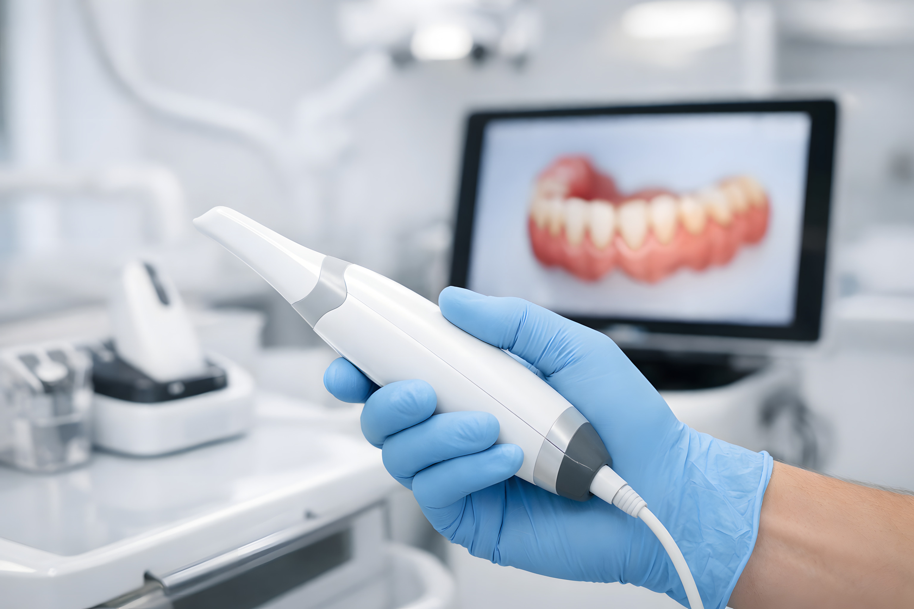
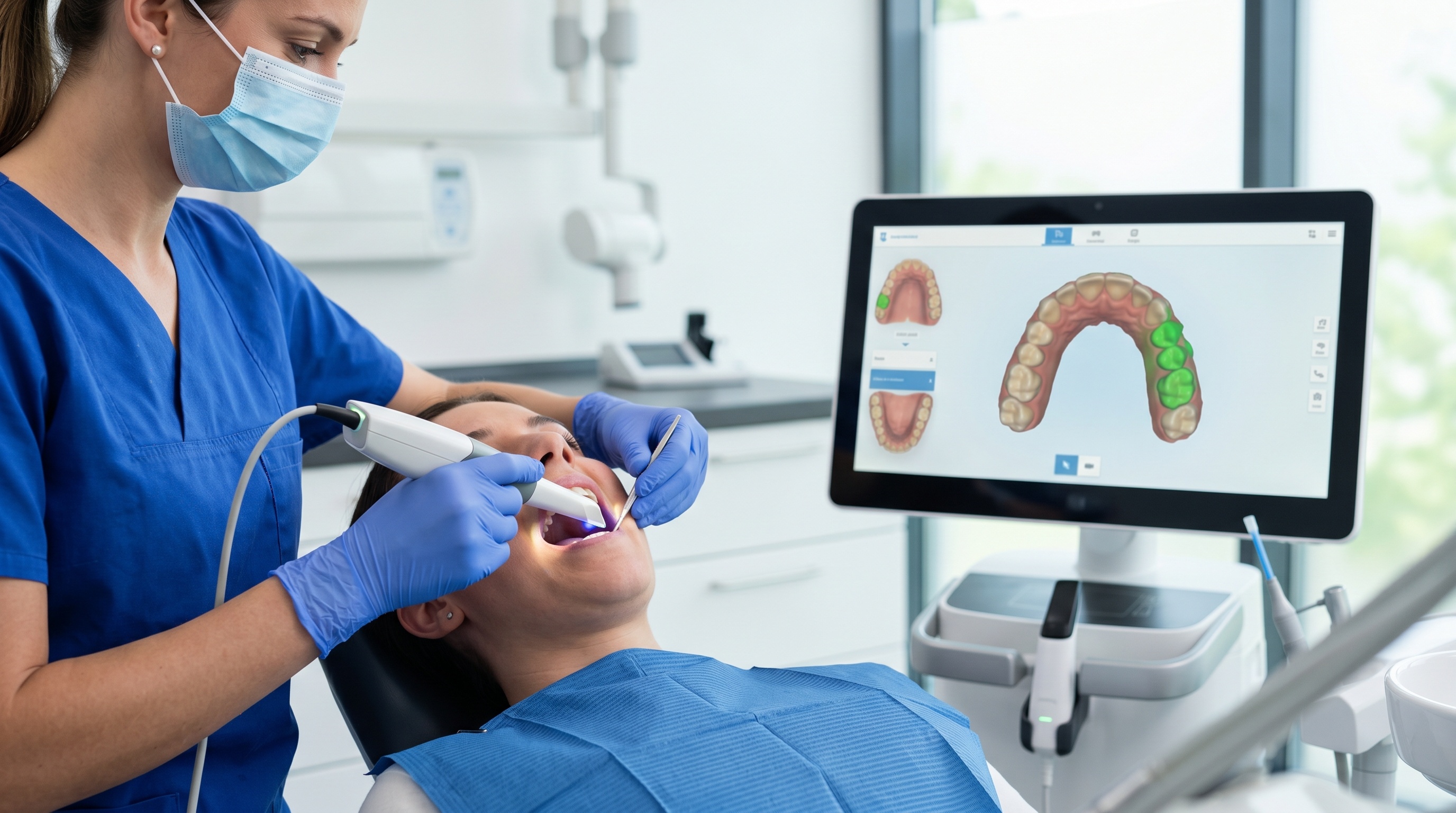
The Medit i900 is the latest generation intraoral scanner from Medit, one of the leading manufacturers of dental scanning technology. It replaces traditional impression trays — the goopy, uncomfortable molds that many patients dread — with a small handheld wand that captures a precise 3D digital model of your teeth and gums in minutes.

The digital scan is more accurate than traditional impressions, more comfortable for the patient, and produces a model that can be used immediately for treatment planning, digital smile design, and fabrication of restorations.

How We Use Digital Scanning

Restorative-driven implant planning

The Medit scan is the first step in digital implant planning. It creates the ideal restoration design first, which is then overlaid onto the CBCT scan to determine exactly where the implant needs to be placed to support it. The surgery follows the restoration plan — not the other way around.

Smile design

The digital model becomes the foundation for designing new tooth shapes, proportions, and arrangements. Dr. Kriston can visualize the proposed smile and create wax-ups or 3D-printed prototypes from the scan data.

Crown, veneer, and restoration fabrication

Digital scans are sent directly to our laboratory electronically, eliminating the shipping time and potential distortion that can occur with physical impressions. The result is restorations that fit with exceptional precision.

Custom Oral Appliances

The same digital scanner that plans your implants and restorations also creates precise models for every custom appliance we fabricate — night guards, TMJ orthotics, sleep apnea devices, sports guards, and whitening trays. A digital scan means a better fit, greater comfort, and no messy impressions.

AI-Assisted Diagnostics and Treatment Planning

Artificial intelligence is transforming how dental diagnoses are made and treatments are planned. At Michael Kriston DDS, AI software is integrated into our digital imaging workflow, providing a powerful second layer of analysis that enhances Dr. Kriston’s clinical judgment.

How We Use AI

Anatomical tracing

AI software within our imaging system can automatically trace critical anatomical structures — such as the inferior alveolar nerve, the maxillary sinus floor, and bone boundaries — on CBCT scans. This provides precise measurements that inform implant placement planning and surgical decisions.

Cavity detection

AI algorithms analyze digital X-rays to identify areas of decay that might be difficult to detect visually, flagging potential cavities for Dr. Kriston’s review. This technology is particularly valuable for detecting early-stage decay between teeth, where cavities are hardest to spot.

Implant treatment planning

AI assists in selecting the ideal implant type and size for a particular site by analyzing the bone volume, density, and anatomy from the CBCT scan. The software can suggest optimal implant dimensions and positioning based on the available bone.

AI as an Assistant, Not a Replacement

Dr. Kriston views AI as a fantastic assistant — but not a replacement for the experienced clinician. AI excels at pattern recognition, measurement precision, and flagging areas of concern. But the final anatomical assessment, the diagnosis, and the treatment plan are always made by Dr. Kriston.

AI doesn’t understand context the way a dentist with 38 years of experience does. It doesn’t know your history, your goals, your bite dynamics, or how this particular finding fits into the bigger picture of your oral health. What AI does is ensure that nothing is missed and that the data Dr. Kriston works from is as comprehensive and precise as possible.

The combination of AI-enhanced imaging and decades of clinical experience is more powerful than either one alone.
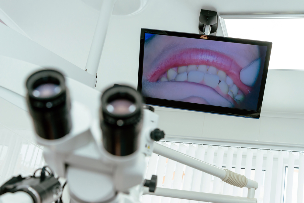
Carestream CS 1500 Intraoral Cameras

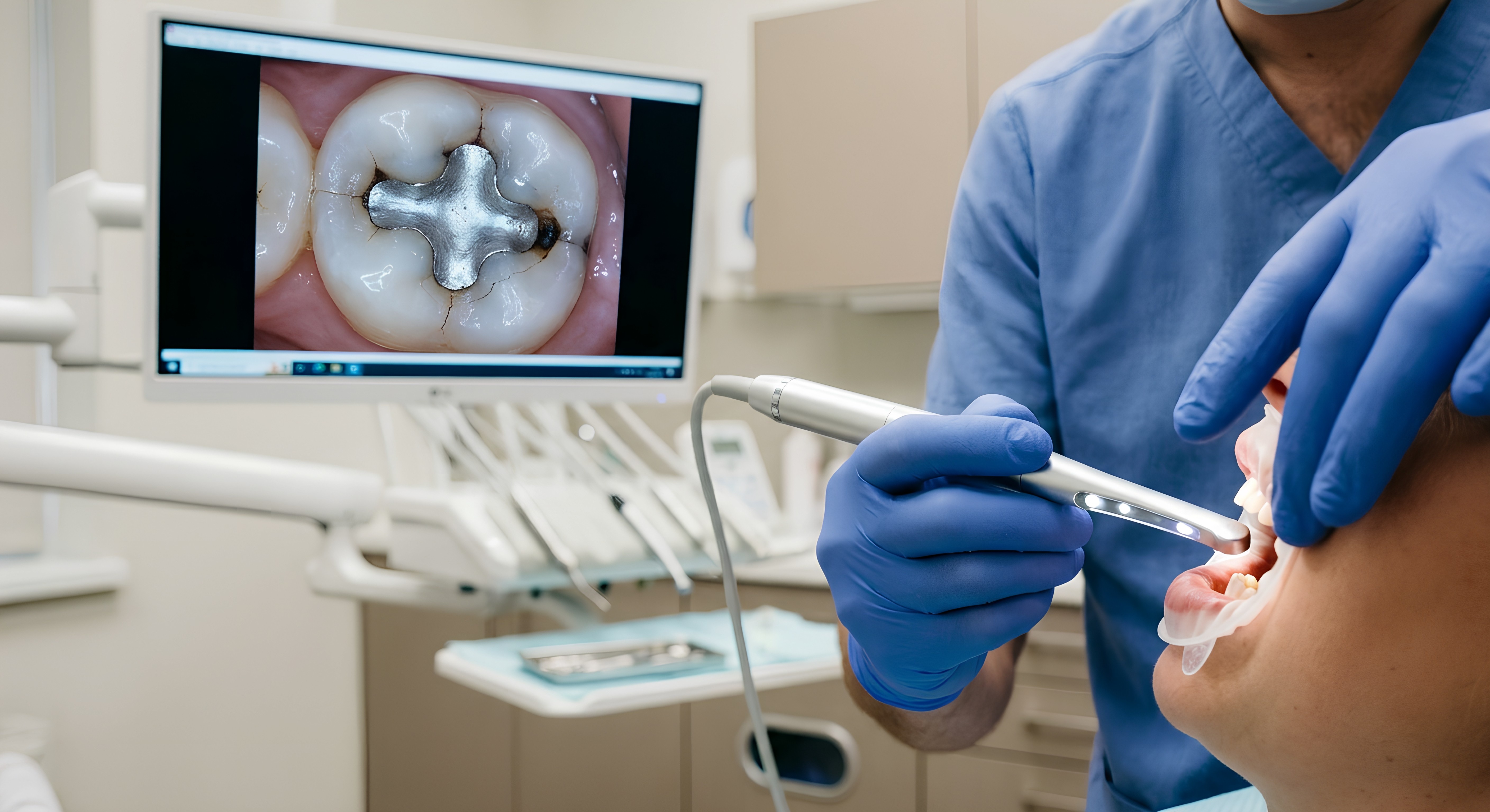
Every treatment room in our office is equipped with a Carestream CS 1500 intraoral camera. These small, pen-sized cameras capture high-resolution images of individual teeth, restorations, gum tissue, and any areas of concern — and display them on a screen in real time so you can see exactly what Dr. Kriston sees.

Intraoral cameras serve multiple purposes in our practice:

Diagnosis

Magnified images reveal cracks, fracture lines, margin breakdown on old restorations, and early signs of decay that may not be visible to the naked eye.

Treatment planning

Detailed images are used alongside X-rays and CBCT scans to evaluate the condition of teeth and plan the most appropriate treatment.

Year-over-year comparison

All intraoral images are stored in your digital patient chart. Dr. Kriston can compare images from year to year to track changes — for example, seeing whether a fracture line in a tooth has extended further, or if new ones have appeared. This longitudinal monitoring helps him intervene at the right time, before a small problem becomes a big one.

Patient education

When you can see what Dr. Kriston sees — a crack, a failing restoration, a change in the gum tissue — the diagnosis and treatment recommendation make immediate sense. No guesswork, no taking our word for it. You see it yourself.

Dexis Digital X-Ray Sensors

Our Dexis digital X-ray sensors capture diagnostic images with significantly less radiation than traditional film X-rays — up to 80% less in some cases. The images appear instantly on screen in high resolution, with no chemical processing, no waiting, and no film waste.

Digital X-rays can be enhanced, magnified, and adjusted for contrast on screen, allowing Dr. Kriston to see subtle details that might be missed on a traditional film. They’re also stored digitally in your patient chart for easy retrieval and comparison over time.

Diolase ST Diode Laser

Our Diolase ST diode laser, manufactured by American Dental Technologies (ADT), is used for all soft tissue procedures in our office, including gum reshaping, gum lifts, crown lengthening, and gingival contouring.

The diode laser offers significant advantages over traditional surgical instruments:

Precision

The laser sculpts tissue at a level of detail measured in fractions of a millimeter — essential for smile design work where gingival zenith positioning must be exact.

Comfort

Laser treatment is gentler on tissue than traditional surgery, with less swelling and less post-operative discomfort.

Sterile contact‍

The laser’s high degree of sterile contact reduces infection risk.

Fast healing

The laser cauterizes as it works, sealing blood vessels and reducing bleeding. This promotes significantly faster recovery compared to conventional surgical techniques.

Digital Photography

Clinical photography is an essential part of diagnosis, cosmetic planning, and treatment documentation at our practice. Dr. Kriston uses digital cameras to capture standardized images of your smile from multiple angles — full face, close-up, profile, retracted views — that serve as the foundation for smile design, facial analysis, and before/after comparison.

These photographs are used for golden proportion analysis, midline assessment, lip line evaluation, and communication with the dental laboratory. They’re stored in your digital chart alongside your X-rays, CBCT scans, and intraoral images — creating a comprehensive visual record of your dental health and treatment history.

All-Digital Patient Records

Every aspect of your care is documented digitally. Your chart, X-rays, CBCT scans, intraoral photographs, digital scans, treatment plans, and clinical notes are all stored in a secure, integrated digital system. This means:
Instant access to your complete dental history at every appointment
Easy comparison of images and records over time for monitoring changes
Secure, encrypted storage that protects your personal health information
Seamless communication with specialists, laboratories, and insurance providers when needed

Sterilization and Infection Control

Patient safety extends beyond diagnosis and treatment. Our sterilization protocols exceed industry standards to ensure that every instrument used in your care is thoroughly cleaned and sterilized.

All instruments are organized in surgical cassettes with surgical wraps — sealed, sterile packages that are opened only at the time of your appointment. Before sterilization, every instrument passes through our Miele Professional G7881 washer — a medical-grade instrument washer that provides standardized, validated cleaning that surpasses hand washing. After washing, instruments are placed in our Midmark autoclave sterilizer, which uses high-pressure steam to achieve complete sterilization.

This two-step process — professional-grade washing followed by autoclave sterilization, with instruments sealed in surgical cassettes — represents the highest standard of infection control in a dental office.

Patient Comfort

Technology isn’t just about diagnosis and treatment — it’s about your experience. We’ve designed every treatment room with your comfort in mind:
Orthopedic support pillows for your back, neck, and legs — because a two-hour appointment shouldn’t mean two hours of discomfort
Warm blankets available for every appointment
iPads with music and video entertainment to help you relax during procedures
Oral conscious sedation available for patients who want an even more relaxed experience
We believe that great dental care should feel as good as it looks. From the technology that makes your treatment more precise to the amenities that make your visit more pleasant, every detail is intentional.

Experience the Difference

If you’d like to see our technology first hand and learn how it can make your dental care more precise, more comfortable, and more predictable, schedule a consultation at our Danville office.

Call (925) 838-2900 or book online. We welcome patients from Danville, San Ramon, Blackhawk, Alamo, Walnut Creek, Dublin, Pleasanton, and throughout the Tri-Valley and East Bay.

Call Us: (925) 838-2900